How to identify varroa mites under a microscope
TL;DR
- Varroa destructor is a reddish-brown, oval mite about 1.1 mm wide and 1.6 mm long.
- You can see it with the naked eye, but 10x to 40x magnification makes it obvious.
- Under a microscope you get eight legs, a flattened crab-shaped body, and piercing mouthparts.
- No other common honey bee parasite matches that profile.
What does a varroa mite actually look like?
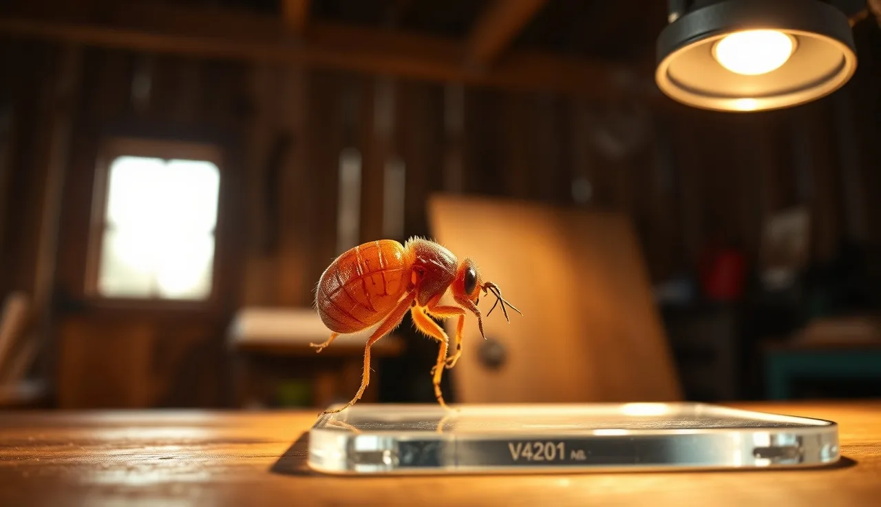
A varroa mite is wider than it is long. That trips people up, because most mites they've seen before are round or stretched out. Varroa is dorso-ventrally flattened, meaning it's squashed top to bottom, and adult females run roughly 1.1 mm wide by 1.6 mm long [1]. Males are smaller and pale, and you almost never see them on adult bees. They spend their whole short lives inside capped brood.
The color is a reddish-brown, close to mahogany. Under bright light you can spot a mature female on a pale-bodied bee with the naked eye, especially on a drone. On a dark bee she hides against the thorax. That's exactly why looking at bees is not a reliable way to monitor mites.
The body surface carries a fine texture of short setae (hairs) that show up under magnification. The cuticle looks leathery, not shiny. Four pairs of legs come off the sides. Eight legs means arachnid, not insect, and that's the marker that settles the whole class question. The legs are short relative to the body and end in small pad-like structures that let the mite grip a bee and slide between abdominal segments.
The mouthparts (the gnathosoma) stick slightly forward from the front edge of the body. They include chelicerae built for piercing and a hypostome that anchors the mite while it feeds. At 40x or higher the chelicerae come into clear view. That structure is what researchers look at to confirm species [2].
What magnification do you need to identify varroa?
Ten-power (10x) is the practical floor. A decent hand lens or the 10x objective on a compound scope will show you the crab-shaped body, the reddish color, and the eight legs. That's usually enough to separate varroa from the other mite genera you might find on honey bees.
Forty-power (40x) is where the call becomes airtight. At that level you can read the gnathosoma shape, the setal pattern on the dorsal shield (the idiosoma), and the leg structure well enough to split V. destructor from its cousin V. jacobsoni (which lives on Apis cerana across parts of Asia and only rarely jumps to A. mellifera) [3]. For a hobbyist in North America, V. jacobsoni isn't a real possibility. It matters if you do formal research or import queens.
A dissecting (stereo) microscope at 10x to 40x is the best tool for this. You set the mite on a white surface or a glass slide, and the low power plus depth of field lets you see the whole body at once without flattening it. Compound microscopes need a proper slide mount to show surface detail, which means preparing the mite, usually in a clearing medium like lactic acid or Hoyer's.
For routine monitoring, a 10x jeweler's loupe or a USB digital microscope in the $30 to $80 range does the job. You're confirming presence, not publishing taxonomy.
How do you collect a mite sample for microscope examination?
The two standard sampling methods are the alcohol wash and the powdered sugar roll [4]. Alcohol wash (70% isopropyl or ethanol) kills and dislodges mites reliably. Sugar is gentler on the bees but shakes fewer mites loose, so it undercounts. If you want a mite for microscope work rather than a population estimate, use the alcohol wash. The mites come out intact.
Collect about 300 adult bees (roughly half a cup) from a brood frame. Take the bees clustered on the frame, not the frame surface, and keep the queen out of the jar. Cover them with enough 70% alcohol to submerge them. Seal and shake for 30 to 60 seconds. Pour the liquid through a fine mesh strainer (150 to 300 micron) into a white tray. The mites drop through; the bees stay on the mesh. Swirl the tray and look for the reddish-brown oval bodies.
To get a mite onto a slide, fish one out of the alcohol with a fine brush or a dissecting needle. Set it dorsal-side up on a glass slide. A drop of 70% alcohol or water holds it in place while you line it up. For a permanent mount, add a coverslip and seal the edges with clear nail polish once the alcohol dries.
Want to study the legs and mouthparts in detail? Clear the mite in 85% lactic acid, heating it for 20 to 30 minutes on a warming plate at around 55 degrees C. The cuticle goes semi-transparent and the internal structures show through [2]. Most beekeepers never need this. It's routine in diagnostic labs.
Always pull your sample from a brood frame. Mites reproduce only in capped brood, so a hive with a low adult-bee count can still carry heavy brood infestation [5]. For the full picture you need both the adult-bee wash and a look inside capped cells.
How do you tell varroa apart from other mites on honey bees?
Three other mite groups turn up in honey bee colonies often enough to cause confusion: Tropilaelaps, Acarapis, and various phoretic hypopus (the non-feeding travel stages of soil mites). Here's how each one differs.
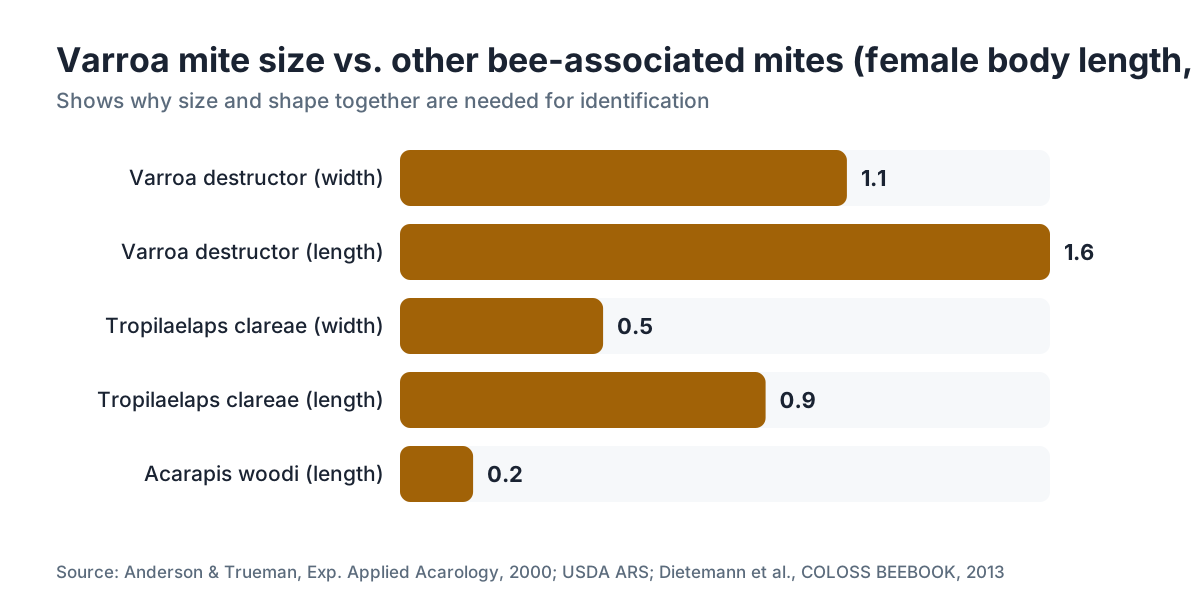
Tropilaelaps (two species, T. clareae and T. mercedesae) are honey bee parasites found in Asia and a serious import concern [6]. Under the scope they look a little like varroa, but they're elongated instead of wider-than-long, smaller (around 0.9 mm by 0.5 mm), and they move fast compared to varroa's slow crawl. Their gnathosoma is long and narrow, not compact. Tropilaelaps is not established in North America or Europe as of mid-2025, but it sits on every biosecurity watchlist.
Acarapis woodi is the tracheal mite. It lives in the prothoracic tracheae of adult bees, so you'll never see it during an external check. Diagnosis means dissecting the thorax and looking at the tracheal tubes under magnification. Healthy tubes are cream or white; infested ones show brown discoloration and mites inside [7]. Size runs about 0.1 to 0.2 mm, ten times smaller than varroa. Adults have eight legs, larvae six, and the diagnostic form is often the six-legged larval stage inside the trachea. If you see large reddish mites on a bee's outer body, it isn't Acarapis.
Hypopus stages of various Acari hitch rides on bees without feeding on them. They carry a distinctive sucking disc (a ventral attachment organ) for clinging to the host. Under magnification the disc jumps out, and the body proportions don't match varroa's flat crab shape.
The shorthand: reddish-brown, wider than long, eight legs, found on adult bee cuticle or in capped brood, in North America or Europe. That's varroa destructor until proven otherwise.
| Feature | Varroa destructor | Tropilaelaps spp. | Acarapis woodi |
|---|---|---|---|
| Size (female) | ~1.1 mm wide, 1.6 mm long | ~0.5 mm wide, 0.9 mm long | ~0.1-0.2 mm |
| Shape | Oval, wider than long | Oval, longer than wide | Elongated |
| Color | Reddish-brown | Pale brown | Pale/cream |
| Location | Adult cuticle, brood | Adult cuticle, brood | Tracheal tubes only |
| Legs (adult) | 8 | 8 | 8 (larvae: 6) |
| Movement | Slow | Very fast | N/A (internal) |
| North American status | Widespread | Absent (2025) | Present but rare |
What do varroa eggs and immature mites look like under magnification?
You only find the full life cycle inside capped brood, so this work means uncapping cells and looking at what's inside. It's standard practice in foulbrood diagnosis labs and university extension diagnostic programs.
The foundress female slips into a cell just before capping, usually a drone cell, because the longer development time gives her more reproductive cycles. Once the cell is capped she lays her first egg, a male, on the cell wall near the base, then female eggs at roughly 30-hour intervals. Male eggs are a bit smaller than female eggs [8]. Under 10x the eggs are white, oval, and about 0.3 mm long. They're easy to lose against the pale wax and larval silk.
Protonymph and deutonymph stages are pale, close to translucent, and look like tiny varroa with unfinished legs. The first nymph has only six leg stumps showing. As they grow they darken toward the reddish-brown adult color. Only mated females that finish development are reproductively viable, and those are the ones that ride out on an adult bee and start the phoretic phase.
Here's something a lot of beekeepers miss: the first female offspring often fails to mature because brood development runs too fast for the mite's cycle. It happens more in worker cells than drone cells, and that's one reason varroa leans hard toward drone brood. Drone brood stays capped about 14 days versus 12 for workers, giving the mite the extra time it needs to finish a full cycle [5].
What equipment do you actually need, and what does it cost?
You don't need a research-grade microscope. Here's the honest rundown.
A 10x loupe or hand lens runs $10 to $30 and is genuinely enough to confirm whether a mite is varroa. Hold it close to your eye, bring the specimen up to the lens, and focus by moving your head. Plenty of experienced beekeepers use exactly this in the field.
A USB or smartphone clip-on digital microscope costs $25 to $100 and puts the image on a screen, which helps when you're showing someone else or want a photo. Resolution swings wildly across that price band. Look for adjustable LED lighting, because the reddish mite body absorbs light in odd ways.
A stereo dissecting microscope in the 10x to 40x range costs $150 to $600 new, less used. This is the tool university extension labs and serious sideliners reach for. Good optics, a working distance long enough to move specimens around, and binocular depth perception that makes the mite's three-dimensional structure far easier to read. AmScope and Motic both make usable entry-level models in that range.
A compound light microscope is overkill for routine varroa ID, but handy if you also want Nosema spore counts, tracheal mite diagnosis, or pollen work from beehive pollen. Budget $300 to $800 for a decent entry model with a mechanical stage.
My honest advice for most hobbyists doing alcohol washes: spend the $25 on a USB microscope and move on. For a sideliner running 20 or more colonies who wants real diagnostics, a used stereo scope earns its keep.
Once you've confirmed an infestation, the free tools at VarroaVault pull treatment thresholds, timing windows, and product picks into one place.
How do you distinguish a live varroa mite from a dead one?
This comes up with alcohol wash counts. After 60 seconds of shaking bees in alcohol, some beekeepers wonder whether the mites they're counting were already dead going in.
Live mites dropped into alcohol stretch their legs briefly before they die. Mites dead for days show retracted legs, duller color than fresh specimens, and sometimes a partial collapse of the cuticle. Under magnification, a mite killed seconds to hours ago looks basically identical to a live one. An old dead mite that has dried out may be shrunken or cracked.
For counting, the distinction barely matters. A dead mite on an adult bee was still an ectoparasite feeding on that bee's fat body. The mite load you measure for treatment decisions counts live and recently dead mites the same way.
One case where it does matter: high numbers of dead mites in a tray or on the bottom board during a natural mite fall check. That tells you something about population size, but it's not a clean number. Natural mite fall swings hard with season and colony behavior, and the Honey Bee Health Coalition's Varroa management guide flatly calls natural mite fall an unreliable standalone monitoring tool [4].
Can you use a phone camera or digital tools instead of a microscope?
Yes, with limits. A modern smartphone with a good macro mode gets you to roughly 5x to 10x effective magnification, enough to see the reddish-brown oval body and count legs. A clip-on macro lens (about $10 to $30) sharpens that and buys you more working distance.
Several beekeeping apps and general image-recognition tools have been tested on varroa photos, with mixed results. A 2021 paper in Computers and Electronics in Agriculture reported a convolutional neural network hitting over 97% accuracy on high-quality images of mites on frames [9]. In the field, with lower image quality, that number drops a lot. These tools aren't ready to replace a wash count, but they help confirm "yes, that reddish thing on this bee is a varroa mite."
For monitoring and treatment decisions, the alcohol wash count expressed as mites per 100 bees is still the standard everyone measures against. The Honey Bee Health Coalition sets a general action threshold of 2 mites per 100 bees (2%) during the brood-rearing season [4], though some extension programs push treatment at 1% in late summer when the colony is raising its winter bees. Identifying the mite and counting the mites are two separate problems that call for different tools.
A solid overview of varroa mite biology and treatment reads well alongside the microscopy details here.
What does varroa damage to bee pupae look like under magnification?
Uncap a cell and the mite's damage shows up. Heavily parasitized pupae carry white fecal spots (mite excrement) on the cell wall and on the pupal body. Under 10x to 20x those spots stand out against the pale background.
The pupae may show deformed, shortened abdomens or crumpled wings if several mites reproduced in one cell. That's the cellular root of deformed wing virus (DWV) transmission, which the mite spreads while feeding. The virus itself doesn't show under a standard light microscope, but the wing deformity in emerged adults is a field sign that mite levels are high enough for multiple mites to share a cell [10].
A single mite in a worker cell usually produces one or two viable female offspring and does less obvious physical damage. Several foundress mites in one cell (which happens at high infestation because more mites compete for brood) drive more deformity and higher reproductive failure.
If you're uncapping brood specifically to gauge how well the mites are reproducing (which tells you whether your mite population is climbing fast or slow), open drone or worker cells at the purple-eye pupal stage and count foundress mites plus viable offspring. A foundress with two or more healthy female offspring counts as successful reproduction. This takes a decent stereo scope and some practice reading developmental stages, but it's genuinely useful for research-minded beekeepers.
Are there microscopy-based tests used in official diagnostic labs?
Yes. State department of agriculture labs and university extension diagnostic services use microscopy for several bee health problems, and varroa confirmation is one of the easier ones. For a submitted sample, a technician runs an alcohol wash, strains the liquid, examines the tray under a stereo microscope, counts mites, and reports the count per 100 bees along with species confirmation.
The USDA Agricultural Research Service Bee Research Laboratory in Beltsville, Maryland is the primary federal reference lab for bee diagnostics in the United States [11]. It accepts submitted samples and can confirm varroa species, identify disease agents, and detect Tropilaelaps (which triggers regulatory action) using morphology and, where needed, molecular methods like PCR.
The National Bee Unit in the UK runs a similar tiered approach: field identification by inspectors, lab confirmation by stereo microscopy, and molecular confirmation for notifiable pests [6].
For North American beekeepers, your state department of agriculture apiarist is the first call for diagnostic help. Most states offer free or low-cost sample submission. If you suspect something odd (not varroa, or varroa acting strangely), submit a sample instead of guessing. Shipping a vial of bees in alcohol costs almost nothing compared to treating the wrong problem.
Extension programs at land-grant universities (Penn State, University of Florida, University of Minnesota, and others) also publish free diagnostic protocols and identification keys you can work from yourself [12].
How do you photograph varroa under a microscope for records or sharing?
Good mite photos help when you're sharing an ID with other beekeepers, submitting to an extension program, or building your own monitoring records over time.
The basic setup: put the mite on a white index card or a glass slide over white paper. Use a strong, angled light source. Don't light straight from overhead, which flattens the image and kills shadow detail. A small LED flashlight held at 45 degrees works well. Get your microscope or macro lens close enough that the mite fills most of the frame.
With a phone, turn on the grid to level the shot and tap the mite body to lock focus. Burst mode helps, since the smallest vibration blurs a macro shot. Fire off 10 frames and keep the sharpest.
With a stereo microscope that has a trinocular head, you can mount a DSLR or mirrorless camera through an adapter. That's the setup university extension labs use for documentation. Exposure is usually manual at ISO 400 to 800, with a mechanical shutter delay to cut vibration.
For asking ID questions online (BeeSource forums, local beekeeper association Facebook groups, your state apiarist's inbox), a clean dorsal view showing the full body next to a common object for scale (a coin, a printed millimeter ruler) beats a blurry close-up every time. The reddish-brown color, the oval wider-than-long shape, and the visible legs are what experienced eyes check when they confirm an ID from a photo.
If you take diagnostics and records seriously, build a simple reference slide collection. One mite per monitoring session, dated and labeled, takes almost no time and gives you a physical log of your colony's history. It also trains your eye. The more mites you look at, the faster you spot them.
Frequently asked questions
Can you see varroa mites without a microscope?
Yes. Adult female varroa run about 1.1 mm wide and 1.6 mm long, visible to the naked eye on a pale-bodied bee or on a white surface after an alcohol wash. But you need at least a 10x loupe to confirm the eight legs, crab-shaped body, and reddish-brown color clearly enough to rule out debris. Naked-eye inspection of adult bees is not a reliable way to monitor mites.
What magnification is best for identifying varroa mites?
Ten-power (10x) is enough to confirm varroa in most situations. Forty-power (40x) shows mouthpart and leg detail, which matters for separating varroa from related species. A stereo dissecting microscope at 10x to 40x is the most practical tool. A USB digital microscope in the $25 to $80 range works fine for routine hobbyist confirmation.
How many legs does a varroa mite have?
Adult varroa mites have eight legs, which puts them with the arachnids (same class as spiders and ticks). Larval varroa have six legs, but you only meet the larval stage inside capped brood cells. The eight-legged adult female on a bee's body is the form most beekeepers see and the one you use for identification.
What color is a varroa mite under a microscope?
Reddish-brown, often called mahogany. The color runs consistent across the dorsal shield (idiosoma) and legs. Males are much paler, nearly white, and smaller than females. The cuticle has a finely textured, leathery look under magnification rather than a shiny surface. Color alone hints at varroa but doesn't confirm it; body shape and leg count finish the job.
How do you tell varroa mites apart from tracheal mites (Acarapis woodi)?
They differ completely in size, location, and look. Varroa females are about 1.1 mm wide and sit on adult bee cuticle or in brood cells. Tracheal mites are 0.1 to 0.2 mm (ten times smaller) and live only inside the thoracic tracheal tubes. You can't see tracheal mites externally at all. Diagnosis means dissecting the thorax and examining tracheal tissue under magnification.
What does a varroa mite look like inside a brood cell?
Inside a capped cell you'll find the reddish-brown foundress female plus her eggs and offspring at various stages. Early nymphs are pale, close to translucent, with unfinished legs. The cell wall usually carries white fecal spots from mite feeding. Under 10x to 20x, those spots and the size gap between foundress and offspring show up clearly.
How do you prepare a varroa mite for microscope examination?
For a quick look, set the mite on a glass slide or white card in a drop of 70% alcohol and view it under a stereo microscope. For a permanent slide, let the alcohol dry, add a coverslip, and seal the edges with clear nail polish. For detailed morphology, clear the mite in 85% lactic acid at 55 degrees C for 20 to 30 minutes to make the cuticle semi-transparent and reveal internal structures.
What is the action threshold for varroa mites, and how is it measured?
The Honey Bee Health Coalition recommends an action threshold of 2 mites per 100 bees (2%) during the brood-rearing season. You measure it with an alcohol wash: 300 bees in 70% isopropyl, shaken 30 to 60 seconds, mites counted in the strained liquid. Late-summer thresholds may drop to 1% because winter bee quality is on the line. Natural mite fall counts aren't reliable for this.
Is Tropilaelaps present in the United States, and how would you tell it from varroa under a microscope?
Tropilaelaps is not established in the United States or Europe as of mid-2025. Under a microscope it differs from varroa by being elongated (longer than wide rather than wider than long), smaller (about 0.9 mm by 0.5 mm), paler brown, and much faster-moving. The gnathosoma is long and narrow versus varroa's compact mouthparts. Report any suspected Tropilaelaps to your state apiarist right away.
Can a smartphone identify varroa mites from a photo?
Smartphone photos with a clip-on macro lens can show enough detail to confirm varroa. AI tools trained on bee images have hit over 97% accuracy on high-quality mite images in controlled settings, but field photo quality drops that number a lot. Photos help confirm 'yes, this is varroa' but don't replace alcohol wash counts for estimating infestation levels and treatment decisions.
Where can you send bee samples for official varroa identification?
The USDA ARS Bee Research Laboratory in Beltsville, Maryland is the primary federal diagnostic reference lab in the US. Most state departments of agriculture also accept submitted samples through their state apiarist programs, often at low or no cost. University extension programs at land-grant institutions (Penn State, University of Florida, University of Minnesota) publish diagnostic protocols and some accept samples.
Do male varroa mites look different from females?
Yes, a lot. Male varroa are smaller, much paler (nearly white or cream), and rarely seen because they never leave the brood cell. Their whole life cycle happens inside capped brood: they mate with the developing female offspring and die in the cell. Uncap brood and you might spot a pale, smaller mite among the darker foundress and her offspring, but males aren't the form you identify from.
How do you count mites per 100 bees after an alcohol wash?
Collect about 300 adult bees from a brood frame into a jar, cover with 70% isopropyl alcohol, seal, and shake for 30 to 60 seconds. Pour through a 150 to 300 micron mesh strainer into a white tray. Count the reddish-brown mites in the liquid. Divide the mite count by the actual bee count (estimate from bee volume, or count the dead bees) and multiply by 100 to get mites per 100 bees.
Sources
- Anderson & Trueman, Experimental and Applied Acarology, 2000, Varroa jacobsoni species description and V. destructor morphometrics: Female V. destructor body dimensions approximately 1.1 mm wide by 1.6 mm long
- Dietemann et al., Journal of Apicultural Research, 2013, COLOSS BEEBOOK Vol. II: Standard methods for Varroa research: Chelicerae morphology and lactic acid clearing protocol for detailed mite identification
- Navajas et al., Proceedings of the Royal Society B, 2010, V. jacobsoni switching to A. mellifera: V. jacobsoni distinguished from V. destructor by gnathosoma and setal pattern at 40x magnification
- Honey Bee Health Coalition, Tools for Varroa Management Guide, 2022 edition: Action threshold of 2 mites per 100 bees; natural mite fall not a reliable standalone monitoring tool; alcohol wash as gold standard
- Rosenkranz, Aumeier & Ziegelmann, Journal of Invertebrate Pathology, 2010, Biology and control of Varroa destructor: Drone brood capped period (~14 days) longer than worker brood (~12 days), explaining mite preference; adult-bee wash and brood examination needed for full picture
- UK National Bee Unit, Tropilaelaps pest alert and identification guide: Tropilaelaps spp. morphology versus varroa; National Bee Diagnostic Centre tiered diagnostic approach
- USDA ARS, Honey Bee Tracheal Mite (Acarapis woodi) information page: Acarapis woodi size (~0.1-0.2 mm), internal location in prothoracic tracheae, brown discoloration of infested tubes
- Garrido & Rosenkranz, Apidologie, 2003, Reproductive biology of V. destructor in worker and drone brood: Male egg laid first, female eggs at ~30-hour intervals; egg size differences visible under magnification
- Babic et al., Computers and Electronics in Agriculture, 2021, CNN-based varroa detection from images: Convolutional neural network achieved over 97% accuracy on high-quality varroa mite images
- Nazzi & Le Conte, PLOS Pathogens, 2016, Ecology of Varroa destructor and DWV interaction: Varroa vectors deformed wing virus during feeding; wing deformity in adults indicates multi-mite cell infestation
- USDA ARS Beltsville Bee Research Laboratory, Diagnostic services: Primary federal reference lab for bee diagnostics in the US; accepts sample submissions for varroa species confirmation and Tropilaelaps detection
- Penn State Extension, Varroa mite identification and management: University extension diagnostic protocols and identification keys available free to beekeepers
- University of Minnesota Bee Lab, Varroa sampling methods: Alcohol wash methodology; late-summer threshold of 1% recommended for winter bee protection
Last updated 2026-07-09