What do varroa mites look like? A visual ID guide
TL;DR
- Varroa destructor is a reddish-brown, crab-shaped external parasite about 1.1 mm wide and 1.6 mm long, roughly the size of a sesame seed.
- Females are visible to the naked eye on adult bees, clinging between abdominal segments.
- Males are smaller, paler, and almost never seen outside capped brood cells.
- You will spot most mites in two places: phoretic on adult bees, and as debris on a sticky board.
What does a varroa mite look like in plain terms?
The adult female varroa mite is reddish-brown to mahogany, oval or slightly kidney-bean shaped, and flattened top to bottom. That flattening is not subtle. The body looks like a disc pressed between two fingers, which is exactly what lets it wedge between the overlapping abdominal plates of a honey bee and hang on through grooming.
Size is the first thing most beekeepers notice. The female measures roughly 1.6 mm long and 1.1 mm wide [1], about the diameter of a sesame seed, and she is consistently wider than she is long. That width-to-length ratio is unusual among mites. It is one of the fastest visual clues that you are looking at Varroa destructor and not some other hive mite. She has eight stubby legs arranged in two rows on the front half of her body, not the rear, because she is an arachnid. There is no clear head-and-neck separation. The capitulum (mouthparts) sticks out from the front but blends into the flat oval when viewed from above.
Color is not perfectly uniform. Freshly molted adults look paler or more orange before the cuticle darkens. Mites dead a while on a sticky board fade toward tan or yellowish-brown. But the live, active female on a bee is unmistakably reddish-brown, and her color pops against the bee's pale intersegmental membrane.
The male is a different animal. He is smaller, pale (off-white to yellowish), and spends his whole life inside capped brood cells. You will almost never see a live male unless you deliberately uncap a pupating cell and go looking. For day-to-day monitoring, the female is the mite you are learning to recognize.
How big is a varroa mite compared to familiar objects?
The female is about 1.1 mm wide, roughly the width of a sesame seed and about one-third the width of a grain of white rice. She is big enough to see with the naked eye against a pale background, which is the whole reason sticky board monitoring works. Numbers help, but comparisons land faster.
Here is how she stacks up:
- Roughly the width of a sesame seed
- About one-third the width of a grain of white rice
- About three times the width of a grain of table salt
- Very close to the width of a standard sewing pinhead
- Clearly visible to unaided eyes in good light, no magnifier required
Many pest mites in agriculture need a hand lens or microscope to spot at all. Varroa does not. That size difference is what keeps field monitoring practical for a hobbyist with a kitchen loupe.
| Object | Approximate size | Comparison to female varroa |
|---|---|---|
| Female varroa (width) | 1.1 mm | Reference point [1] |
| Sesame seed (width) | ~1.0-1.5 mm | About the same |
| Grain of table salt | ~0.3 mm | Varroa is ~3x wider |
| Pinhead (standard sewing pin) | ~1.4 mm | Very close |
| Worker bee front leg at tibia | ~0.5 mm | Varroa is about twice as wide |
Under a 10x hand lens or a phone macro lens, the mite gets dramatically easier to read. You can make out the leg arrangement, the flat body profile, and the color difference between the dorsal (top) and ventral (bottom) surface. If you are training your eye, ten minutes with a sticky board sample and a loupe is the fastest education money can buy [10].
What do varroa mites look like on a bee?
On a living worker or drone, the mite almost always parks between the abdominal tergites, the overlapping hard plates on the bee's back. She sits with the long axis of her body running across the bee's width, not along it, another artifact of her wider-than-long shape. From above she looks like a small brown button tucked into the seam between two segments.
The most reliable place to look is the soft intersegmental membrane between segments 3 and 4 on the abdomen. That area gives the mite access to the bee's fat body tissue, which is what she feeds on. Mites shift position, though, and have turned up on the thorax and near the head on badly infested bees. The color contrast is best when the bee holds still: reddish-brown mite against pale yellowish membrane.
Drones often carry more mites per bee than workers do. Drone brood cells are preferred reproductive sites, and drones drift between colonies more freely, so their phoretic loads run higher [2]. If you run a wash count and your sample is mostly drones, your result may overstate the colony average.
On a dead or chilled bee laid on a white paper towel, the mites often drop off or become easy to see because the bee stops grooming. That is why researchers sometimes chill samples before examination. For field ID, brushing live bees onto a white surface, or doing an alcohol wash and straining the liquid through a coffee filter, gives you mites you can study up close with a hand lens.
What do varroa mites look like in brood cells?
Inside a capped cell, the reproductive life of varroa becomes visible, and it is genuinely strange to see the first time. A foundress female enters the cell just before capping and hides in the brood food at the bottom. After capping she climbs onto the developing pupa and lays eggs on a timed schedule. The first egg usually becomes a male, and the eggs that follow become females [3].
Uncap a cell and you may see three things at once: the original foundress (dark reddish-brown), one or more daughter mites mid-molt (pale, soft-bodied, legs not fully formed), and the male (small, rounded, pale). The foundress is easy to pick out by her adult color. Immature females pass through protonymph and deutonymph stages that look more like the adult over time but stay whitish or cream until the final molt.
The Honey Bee Health Coalition's Varroa management guide notes that worker brood needs about 12 days from capping to emergence while drone brood needs 14 to 15 days [4]. That extra time in drone brood lets more daughter mites finish developing and mate per cycle, which is why drone trapping works as a control tool.
To see reproductive mites yourself, look for capped cells with a slightly sunken or punctured cap, though plenty of infested cells look perfectly normal from outside. Uncap 50 to 100 cells from the same age cohort with a capping scratcher, then use a bright light and a hand lens on the pupae and cell bottoms. You may need to gently roll or lift the pupa out to see mites clinging to the body.
What do varroa mites look like on a sticky board?
On a sticky board, varroa show up as small reddish-brown to brownish-orange oval specks, usually 1 mm or a touch smaller, lying flat against the surface because their bodies are flat. A sticky board (also called a sticky sheet or IPM bottom board insert) collects natural mite fall: mites that drop off bees or emerge from brood and drop through the mesh screen above. Counting them over 24 or 72 hours gives you a natural fall rate that loosely tracks infestation, though the relationship is noisy enough that most researchers now prefer an alcohol wash or sugar roll for counting [5].
Under a hand lens, the leg arrangement shows on fresh specimens, and you can confirm the kidney-bean outline. The trouble is background noise. A sticky board also collects wax flakes, pollen granules, cocoon fragments, and propolis bits, some of them close to varroa in color and size. Here is a field guide to sorting them out [10]:
| What you see | Color | Shape | Legs visible? | Most likely ID |
|---|---|---|---|---|
| Reddish-brown oval, 1 mm | Reddish-brown | Oval, wider than long | Yes under lens | Varroa female |
| Pale oval, <0.8 mm | Off-white/cream | Rounder | Yes under lens | Varroa male or immature |
| Brownish angular fragment | Variable brown | Irregular | No | Wax or propolis |
| Yellow-orange granule, round | Yellow to orange | Spherical | No | Pollen grain |
| White oval flake | White | Flat, irregular edge | No | Wax capping fragment |
| Tiny dark oval, <0.5 mm | Dark brown/black | Oval | Barely visible | Pollen mite or debris |
The Honey Bee Health Coalition treats a natural fall of roughly 2 to 4 mites per day as a prompt to run a more precise wash count, not as a standalone treatment trigger [4]. Do not shrug off one mite a day in late summer. Seasonal population dynamics mean low fall in July can reach explosive levels by September without action.
To count faster, slide the board under a bright lamp, use a 10x loupe, and work in grid sections across the board. Mark each mite with a fine-tipped Sharpie as you go so you never count it twice.
How do varroa mites move and behave on a bee?
Phoretic mites (the ones riding adult bees between reproductive cycles) are not passive hitchhikers. They move between bees during trophallaxis and grooming, and they read chemical cues from brood cells to time their jump from nurse bee to larva just before the cell is capped.
Watch a heavily infested bee under magnification and you can sometimes see the mite pivot and reposition. Her legs have adhesive pads (pulvilli) that grip bee cuticle and hair hard, which is exactly why bees struggle to groom them off. Colonies that groom aggressively do remove and damage more mites, and breeding for hygienic behavior or varroa-sensitive hygiene (VSH) targets that mechanism directly [6].
Mites are easiest to see when the bee is chilled, freshly emerged, or heavily loaded. A bee carrying three or more mites jumps out far more than one carrying a single mite half-hidden under a tergite. If you are doing a visual inspection instead of a wash count, look at the underside of the abdomen and the joints between segments, not the top of the thorax.
What distinguishes varroa from other mites found in hives?
A flat, reddish-brown, sesame-seed-sized oval on an adult bee or sticky board in North America, Europe, Australia, or New Zealand is almost certainly Varroa destructor. No other common hive mite matches that exact mix of color, size, and shape. Several other species do show up, and mixing them up leads to real management errors.
Tropilaelaps species are the closest look-alikes by ecological role, but they are smaller (about 0.9 mm long), more elongated, reddish-brown, and move much faster across a surface. They are not established in North America or Europe as of this writing [7]. If you keep bees in Asia, Tropilaelaps is a serious problem.
Acarapis woodi, the tracheal mite, is invisible without a microscope. It lives in the breathing tubes of adult bees, so you will never see it on a sticky board or on a bee's surface with any normal inspection. Detecting it takes a completely different method.
Hypoaspis and other predatory mesostigmatid mites turn up on sticky boards now and then. They are generally elongated, faster-moving when alive, and smaller than varroa females.
Pollen mites (family Acaridae) are small, pale, and round. Nothing about them reads like varroa.
How do you confirm a varroa ID if you are not sure?
For most monitoring, a visual ID off a sticky board or a bee is reliable once you have seen a confirmed mite a few times. When you want certainty, an alcohol wash settles it. Place roughly 300 adult bees (about half a cup by volume, using nurse bees off a brood frame) into a jar with 70% isopropyl alcohol, shake for 60 seconds, and strain through a mesh into a white container with water. Mites sink and are easy to count and examine [5].
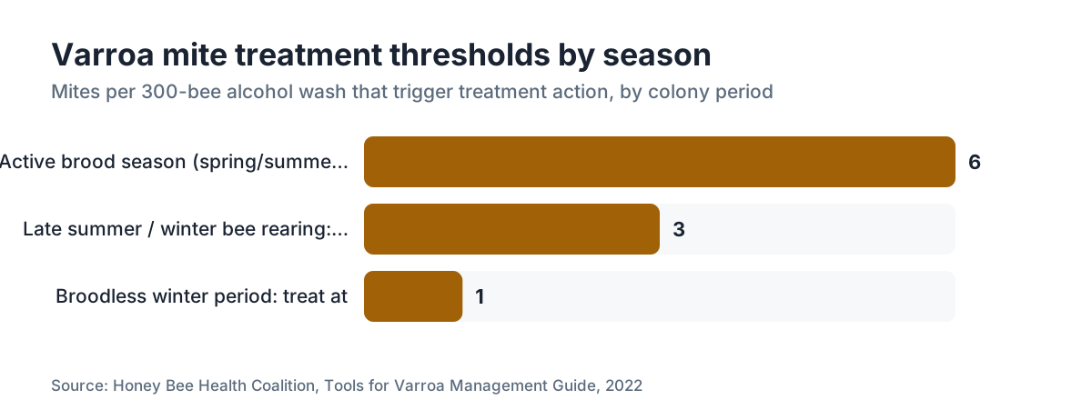
The Honey Bee Health Coalition and many state extension services recommend treating when counts hit 2% to 3% of the bee sample, which is 6 to 9 mites per 300-bee wash, during the active season [4].
Microscopy is not necessary for species-level ID in regions where varroa is the only large bee-parasitic mite. But if you want to study the anatomy, any 40x stereo dissecting microscope shows the full external morphology. University extension labs will often identify submitted specimens when you are genuinely stuck.
If you want a system for keeping track, tools at VarroaVault organize sticky board, alcohol wash, and brood inspection records so you can watch infestation trends over time instead of reacting to one data point.
For the full picture on biology, management options, and treatment thresholds, the varroa mite hub page covers it. For inspection and treatment gear, a comparison of beekeeping supply companies and free shipping honey bee supply companies can help you equip yourself without overspending.
What does varroa mite damage look like on bees?
Spotting mites is one skill. Spotting mite damage is another, and both matter. The clearest signal is deformed wing virus (DWV) in adult bees. Varroa transmits DWV while feeding on fat body tissue inside capped cells, and heavily infested colonies push out a recognizable share of adults with shriveled, stubby, or crumpled wings. Those bees crawl at the entrance, cannot fly, and die fast. See even a handful of crumple-winged bees in a single day and mite pressure is probably already serious [8].
Abdominal shortening is another documented symptom. Bees that developed under heavy parasitism can end up measurably shorter in the abdomen, reflecting fat body loss during the pupal stage. That one is hard to notice without a side-by-side comparison.
Pupae in uncapped cells may show gray or brownish spotting where mites fed. This can look like other brood diseases, so do not diagnose European or American foulbrood off mite damage alone. If you are unsure about brood disease, send a sample to your state apiary inspector.
Colony-level signs of heavy infestation include spotty brood (as infested brood dies and gets pulled by hygienic bees), sudden collapse in late summer or fall, and what beekeepers call fall dwindle: a colony shrinking fast with no obvious pesticide or queen event. None of these are diagnostic alone. Paired with a high alcohol wash count, they tell a coherent story.
When can you see varroa mites most easily during the season?
Mites are present year-round in temperate climates, but how easily you see them shifts with the calendar. The easiest windows are the broodless ones, when nearly every mite is riding an adult bee.
In late winter and early spring, colonies have little or no brood, so almost all mites are in the phoretic phase on adults. This is one of the best times to confirm mites visually by examining bees, and it is also when oxalic acid vaporization works best, since few or no mites are hiding inside capped cells [9].
Through spring and summer, the mite population grows roughly in step with capped brood. As brood ramps up, mites vanish into cells and phoretic counts look low, which can trick you into thinking you have no problem. Cornell's New York State IPM Program notes that mite populations can more than double between July and September as brood peaks and late-season bees are reared [11].
In late summer and early fall, mite numbers peak and are most visible. Workers carrying multiple mites, crawling bees with deformed wings, and high sticky board counts tend to hit together in August and September in the northern hemisphere. This is the highest-stakes monitoring window of the year, because colonies going into winter with heavy mite loads have poor survival odds.
Winter, in climates with a true broodless stretch, gives you one more detection window. Natural fall is easy to count on a sticky board because baseline debris is low, and phoretic mites on bees sit at their most exposed.
How do you photograph varroa mites for documentation?
A clear photo is useful for your state apiary inspector, for training other beekeepers, and for tracking evidence over time. The simplest method wins: do an alcohol wash, pour the strained liquid into a shallow white dish in bright outdoor light or under a daylight-balanced lamp, and shoot straight down with your phone in portrait or macro mode. Mites sink and stand out against the white. Even a basic smartphone produces images good enough to count and share.
For close-up structural shots, set a few mites on white card, let the alcohol evaporate so they lie flat, and shoot with a clip-on macro lens (under $15 at most electronics retailers). At 10 to 15x macro, the leg arrangement, body outline, and color all read clearly.
For sticky board documentation, photograph the whole board in even light first for the overall count, then use a macro lens on any clusters of mite-like objects to confirm identity.
State apiary inspectors and university extension entomologists can often confirm ID from a clear photograph. If you work with a local beekeeping association, a reference set showing mites at different life stages, on bees, in cells, and on sticky board material, is genuinely useful for group training.
Frequently asked questions
What do varroa mites look like on a sticky board?
On a sticky board, varroa females appear as small reddish-brown to mahogany oval specks, roughly 1 mm across, lying flat against the surface. Under a 10x loupe, you can see the wider-than-long kidney-bean shape and stubby legs. Common lookalikes include wax flakes (irregular, white), pollen grains (round, yellow-orange), and propolis fragments (darker, irregular). Mites are distinctly oval and symmetrical compared to all of these.
Can you see varroa mites with the naked eye?
Yes. Adult female varroa mites are about 1.1 mm wide, just large enough to see without magnification against a pale background in good light. On a white sticky board or a white dish after an alcohol wash, they show as tiny brownish oval dots. On a dark-colored bee in a hive, they are much harder to spot without a hand lens. A 10x loupe makes confirmation quick and confident.
What color are varroa mites?
Adult female varroa mites are reddish-brown to mahogany. The shade varies slightly: freshly molted females run more orange, and older or dead mites fade toward tan. Males are much paler, off-white to yellowish, and are almost never seen outside capped brood cells. Immature mites in the protonymph and deutonymph stages are cream or whitish until their final molt hardens and darkens the cuticle.
How small is a varroa mite exactly?
The female is about 1.6 mm long and 1.1 mm wide, making her wider than she is long. That unusual ratio is one of the fastest ID clues. The male is smaller, roughly 0.7 to 0.8 mm, pale, and stays inside capped cells. For a practical reference, the female is close to the width of a sesame seed and about one-third the length of a grain of rice.
Do varroa mites move fast?
Not particularly. Phoretic female varroa mites move at a slow, deliberate pace next to many other mite species. Under a microscope or hand lens, you can watch them reposition on a bee, but they do not scatter fast when disturbed the way some predatory mites do. That slow movement, plus sticky adhesive pads on their legs, is part of why bees have such a hard time grooming them off.
What do varroa mites look like inside a brood cell?
Inside a capped cell, the foundress female is the same reddish-brown as her phoretic form. Her offspring pass through pale, soft-bodied protonymph and deutonymph stages that look cream or whitish against the pupal bee. The male is small, rounded, and pale. To see them, uncap a capped worker or drone cell with a capping scratcher and examine the pupa and cell bottom under bright light with a hand lens.
How do I tell a varroa mite apart from other hive mites?
Varroa females are reddish-brown, distinctly wider than long, and about 1.1 mm wide. No other common North American or European hive mite matches that combination. Tracheal mites (Acarapis woodi) are microscopic and invisible without dissection. Pollen mites are pale and round. A flat, reddish-brown, sesame-seed-sized oval on a bee or sticky board in North America, Europe, Australia, or New Zealand is almost certainly Varroa destructor.
What does varroa mite damage look like on adult bees?
The clearest sign is deformed wing virus (DWV): wings that are shriveled, stubby, or crumpled rather than full-length. Affected bees crawl at the hive entrance and cannot fly. Heavily parasitized bees may also have shortened abdomens. At the colony level, mite damage shows as spotty brood (from hygienic removal of infested cells), population collapse in late summer or fall, and a higher-than-normal share of bees unable to fly.
Is a hand lens enough to identify varroa, or do I need a microscope?
A 10x hand lens or loupe is enough to confirm varroa in the field. You can see the oval outline, the reddish-brown color, the flattened body, and on clear specimens, the leg arrangement. A microscope adds confidence for beginners or for distinguishing damaged specimens, but it is not necessary for routine monitoring in regions where Varroa destructor is the only large bee-parasitic mite present.
At what mite level should I be concerned?
The Honey Bee Health Coalition recommends treating when alcohol wash counts reach 2% (about 6 mites per 300-bee sample) during the active brood season, and 1% (3 mites per 300 bees) in late summer when winter bees are being raised. Sticky board counts of 2 to 4 mites per day are a prompt to run a more accurate wash count, not a standalone threshold. Natural mite fall alone is too imprecise for treatment decisions.
Do varroa mites look different in different seasons?
The mites themselves do not change appearance seasonally, but where and how you see them does. In late winter broodless periods, nearly all mites are phoretic on adult bees, so visual detection on bees is easiest. During peak brood season in spring and summer, most mites hide inside capped cells and phoretic counts drop. In late summer, mite populations peak and mites on adult bees are most numerous and visible.
Can varroa mites infest other bee species?
Varroa destructor originally parasitized Apis cerana (the Asian honey bee) and jumped to Apis mellifera (the European honey bee) in the mid-20th century. It can also infest Apis dorsata under experimental conditions. It does not establish on bumble bees, solitary bees, or other non-Apis species. If you keep Apis mellifera, you are the primary audience for varroa management; beekeepers working with other species face a very different parasite profile.
How do I photograph varroa mites to send to my state inspector?
Do an alcohol wash, strain the liquid into a shallow white dish, and shoot straight down in bright light with your phone's macro or portrait mode. Mites sink and appear as clear brownish ovals against the white. For closer detail, a clip-on macro lens for smartphones works well. Most state apiary inspectors can confirm varroa ID from a clear phone photograph without needing a physical specimen.
Sources
- USDA Agricultural Research Service, Varroa destructor biology page: Female Varroa destructor measures approximately 1.6 mm long and 1.1 mm wide, wider than long, with a flattened oval body
- University of Minnesota Extension, Varroa mite page: Drone brood cells are preferred by varroa for reproduction, and drone bees carry higher phoretic mite loads
- Honey Bee Health Coalition, Tools for Varroa Management Guide (2022 ed.): The foundress female varroa lays her first egg as a male and subsequent eggs as females on a timed schedule after cell capping
- Honey Bee Health Coalition, Tools for Varroa Management Guide (2022 ed.): Recommended treatment threshold is 2% mite load (approximately 6 mites per 300-bee alcohol wash) during brood season; worker brood capping to emergence takes about 12 days, drone brood 14-15 days; natural mite fall of 2-4 mites/day warrants a follow-up wash count
- Penn State Extension, Varroa mite sampling methods: Alcohol wash is the gold standard for mite counting; approximately 300 nurse bees (half-cup volume) are washed in 70% isopropyl alcohol and strained for counting
- USDA ARS Baton Rouge Bee Lab, Varroa-sensitive hygiene (VSH) research: Varroa-sensitive hygiene (VSH) bee stocks detect and remove mite-infested pupae, significantly reducing mite reproduction in colony
- University of Florida IFAS Extension, Varroa mites and deformed wing virus: Varroa mites transmit deformed wing virus (DWV) during feeding on bee fat body tissue in capped cells, resulting in adult bees with shriveled or crumpled wings
- EPA, Oxalic acid product registration and label guidance for bee hive use: Oxalic acid vaporization is most effective during broodless or low-brood periods when most mites are in the phoretic phase on adult bees
- Ohio State University Extension, Varroa mite identification and monitoring: Visual ID of varroa on sticky boards requires distinguishing reddish-brown oval mites from wax flakes, pollen, and propolis debris; a 10x hand lens is sufficient for field confirmation
- Cornell University, New York State IPM Program, Varroa mite management: Mite populations can more than double between July and September as brood production peaks and late-season bees are reared
Last updated 2026-07-09