Pictures of varroa mites: what you're actually looking at
TL;DR
- Varroa destructor is a reddish-brown, oval mite about 1.1 mm wide and 1.6 mm long.
- You can see it with the naked eye, but it hides easily in a busy hive.
- It looks like a tiny crab clamped between the bee's abdominal plates.
- Knowing what you're seeing, and where to look, is where any real mite plan starts.
What does a varroa mite actually look like?
Varroa destructor is reddish-brown and shaped like a flattened oval, wider than it is long. An adult female measures roughly 1.1 mm wide by 1.6 mm long [1]. That's smaller than a sesame seed. But it's big enough to see with your eyes alone if you know where to look and you have decent light. The males are smaller, paler, and you'll almost never see one, because they stay in the brood cell and die soon after mating.
The body surface is covered with short, stiff hairs (setae) that help the mite grip the bee's cuticle. Under a loupe or dissecting microscope, those hairs are obvious, and the eight legs are clearly visible. Eight legs put varroa in the class Arachnida, alongside spiders and ticks, not among the six-legged insects [2]. That taxonomy matters at the treatment level: the chemistry that kills a mite targets arachnid biology and does not hit a bee the same way.
Color runs from pale orange-brown in a freshly matured mite to deep reddish-brown in an older one. On a light thorax, a mite can look almost rust-red. On the dark cuticle of an old forager, you can walk right past it. That contrast, or the lack of it, is exactly why looking with your eyes is a bad way to measure how many mites you actually have.
How big is a varroa mite compared to a honey bee?
A worker honey bee runs about 15 mm long. The mite at 1.1 mm wide is roughly 7 percent of the bee's body width. That number sounds tame until you scale it up. Proportionally it's like a creature the size of a dinner plate clamped to a person's back [2]. Extension programs reach for that image again and again because it makes the parasite load land for a new beekeeper.
Here are the plain numbers side by side:
| | Length | Width |
|---|---|---|
| Adult female varroa | 1.6 mm | 1.1 mm |
| Adult male varroa | ~0.8 mm | ~0.7 mm |
| Worker honey bee | ~15 mm | ~5 mm |
| Varroa as % of bee length | ~11% | ~22% |
A macro lens or an electron microscope lies to you about scale. A sharp 10:1 macro shot makes the mite look the size of a golf ball. New beekeepers see those photos and assume they'll spot a mite instantly. Then they look at a real frame packed with bees, and the mite is a small reddish speck moving fast [1].
Where exactly do you see varroa mites on a bee?
The mite favors the soft membrane between the bee's abdominal tergites, the overlapping plates across the top of the abdomen. It pierces the membrane there and feeds on fat body tissue. A 2019 study in Scientific Reports (Ramsey et al.) settled the feeding question after decades of assumption. As the paper states, varroa "primarily consumes fat body tissue rather than hemolymph" [3].
When you're looking at a live bee, check the underside of the abdomen first, between segments three and four, and four and five. Mites also turn up near the thorax, under the wing bases, or at the head-thorax junction. A mite riding on top of the thorax between the wings is the position you see in most field photos, and the easiest to catch by eye.
On emerging brood, a freshly eclosed bee sometimes carries mites still finishing their cycle and not yet tucked into position. You'll see them on the pale, soft cuticle before the bee darkens. That's one reason uncapping capped brood tells you so much, even though it takes more work than an alcohol wash or sugar roll.
Nymphs, the immature stages, are white to pale yellow and much harder to catch. They sit inside brood cells during the reproductive cycle. To find them, uncap suspect cells (sunken or discolored caps are a starting point) and look with a loupe.
What do varroa mites look like on a sticky board?
A sticky board under a screened bottom board catches mites that drop off bees over a 24 or 48 hour window. On the board they look like tiny reddish-brown ovals scattered across the white or grid-marked surface. Many land upside down (legs up), so you're looking at the pale tan underside and eight stubby legs.
Debris fools people. Pollen grains, wax flakes, and small bits of propolis all fall to the board, and some run close to mite size. Here's the tell: mites are uniformly oval and flat, they don't taper to a point, and their segmented legs show clearly under a 10x loupe. Pollen grains are usually more symmetrical, with surface texture that reads different from the smooth-to-hairy mite cuticle.
The Honey Bee Health Coalition's "Tools for Varroa Management" guide lists a 24-hour natural mite drop count as one monitoring option [4]. A drop of 0 to 1 mites per day reads low-risk. Counts above 10 per 24 hours in mid-summer usually mean the colony is already past the treatment threshold. Those are rules of thumb, not law, and the sticky board undercounts real infestation next to an alcohol wash. Treat it as a trend tool.
Want an accurate photo of board mites? Shoot in good natural light with your phone's macro mode, or clip on a macro lens. A 10x loupe with a built-in LED handles field ID fine.
What do varroa mites look like in brood cells?
Inside a capped cell, the whole reproductive cycle is invisible unless you uncap and look. The foundress female slips into the cell about 20 hours before it's capped, hides under the larval food, and starts laying once the cell seals. The first egg makes a male. The eggs after it make females.
Uncap a cell and you'll usually find the reddish-brown adult foundress plus one or more pale, soft-bodied nymphs clinging to the pupa. The nymphs are translucent white to pale yellow. Early on the pupa can look normal. But heavily parasitized pupae show stubby, deformed abdomens and malformed wings from Deformed Wing Virus (DWV), which varroa moves into the colony [5].
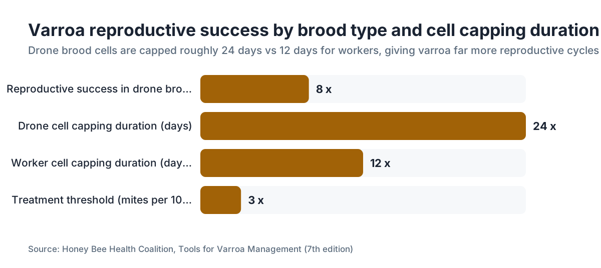
Drone brood is your best window. Varroa reproduces about 8 times more successfully in drone brood than in worker brood, because drone cells stay capped longer (roughly 24 days versus 12 for workers) and that gives the mite more cycles [4]. Pull a drone frame and open cells with a capping scratcher or an uncapping fork. Drop the pupae into a jar of water or alcohol. Any mites float up or stand out against the pupa's body.
Brood-stage photos taken with a digital scope or a good macro lens on a DSLR show the full scene: white nymphs on the pupa, the foundress in place, the deformation already setting in. If a local university or extension lab has a dissecting microscope, ask to look at an infested brood sample. You don't forget it.
How do you photograph varroa mites yourself?
You don't need a scanning electron microscope. A smartphone plus a $10 to $30 clip-on macro lens gets you a solid photo of varroa on a bee, and so does a dedicated macro lens on a mirrorless or DSLR body.
For bees on a frame: chill the frame briefly (a few minutes in a cooler, not a freezer) to slow the bees. That buys you a few seconds of stillness to spot and shoot. Look at single bees under direct sun or a headlamp set to throw raking light across the abdomen. Raking light (a low angle, skimming the surface) picks up the mite's three-dimensional shape far better than flat overhead light.
For alcohol wash samples: pour the gray liquid through a fine mesh strainer over a white tray after the wash. Mites, bee legs, and debris collect on the mesh. Rinse gently. The mites read as reddish-brown ovals. On white, in good light, a phone in macro mode gets a usable shot.
Scanning electron microscope (SEM) images, the kind in journal papers and university fact sheets, show everything: the setae, the mouthparts (chelicerae and pedipalps), the leg joints. These are the images beekeeping books reprint. Penn State Extension and University of Florida IFAS have published some of the most-cited SEM and light-scope images in their varroa materials [6][7].
To go further, a digital USB microscope (roughly $30 to $80 from electronics sellers) magnifies a sample 40 to 200x and saves straight to a computer. Handy for anyone taking mite ID and monitoring seriously.
How can you tell varroa apart from other hive pests by sight?
A handful of other hive organisms get mistaken for varroa, especially by someone looking at a magnified image for the first time.
Tracheal mites (Acarapis woodi) run about 0.14 to 0.18 mm and live inside the bee's tracheal tubes, so you will never see one by eye or even with a hand loupe. If someone says they spotted tracheal mites in the hive with the naked eye, they're looking at something else.
Pseudoscorpions show up in hives now and then, and they're on your side, eating small hive beetles and varroa. They're brown, they carry big pincer-like pedipalps, and they move fast. Once you've seen both, they look nothing like a mite. A beginner might still flag a small one as a parasite.
Pollen mites (Winterschmidtiidae and related families) turn up in stored pollen. They're smaller than varroa, often pale or translucent, and they lack the flat oval shape.
Small hive beetle (Aethina tumida) larvae are white and segmented, nothing like varroa's flat oval. The adult beetles are small, dark, and hard-shelled, also nothing like a mite.
The cleanest varroa ID is the full combination: reddish-brown, oval, flattened, about 1.1 mm wide, eight legs, on or near the abdomen. Nothing else in a hive checks every box. If you're stuck, a university apiary diagnostic lab confirms it from a sample. Many state agriculture departments run this free or cheap for registered beekeepers.
What does a heavy varroa infestation look like at the hive level?
One mite is small and easy to miss. A heavy infestation is loud. When levels climb, the hive throws signs you can read without a magnifier.
The first thing you notice is bees with deformed wings: crumpled, stubby, or shrunken wings that can't fly. That's Deformed Wing Virus, which varroa carries into the bee during feeding and larval development [5]. A hive where 5 percent or more of emerging bees show deformed wings is in real trouble. By the time deformed wings are common, mite levels are usually well past threshold.
You'll also see "crawlers," bees that can't fly and wander the bottom board or landing board. Some have deformed wings. Others carry neurological damage from DWV or other pathogens varroa moves around.
The brood pattern falls apart too. Cells sit empty, brood dies, and the population collapses through what researchers call parasitic mite syndrome: sunken and perforated cappings, scattered brood, secondary bacterial infections. A colony crashing from varroa can look like American foulbrood to a beginner, which is one more reason a real diagnosis matters.
For counting methods and treatment timing, varroa mite management gets full treatment elsewhere on this site. Nailing the visual ID is step one. Knowing what to do next is step two.
The Honey Bee Health Coalition, whose "Tools for Varroa Management" guide is the most-used practical reference in the U.S., sets the treatment threshold at 3 percent (3 mites per 100 bees) during the brood-rearing season [4]. Above that line, a colony is statistically likely to collapse before winter without help.
What do published varroa mite photographs actually show, and where do they come from?
The images in research papers, extension bulletins, and beekeeping books come from a short list of sources: scanning electron microscopy done at university and federal labs (USDA-ARS, University of Florida, Cornell), light-scope shots from diagnostic samples, and field macro photos from researchers and beekeepers.
The USDA Agricultural Research Service Beltsville Bee Lab has produced SEM images of varroa that show up everywhere, from EPA pesticide registration files to state apiary training packets [8]. They're usually public domain, because federal funding paid for them.
University of Florida IFAS Extension keeps a varroa fact sheet (updated now and then) with both SEM images and practical field ID photos [7]. Penn State Extension's bee materials run just as well-illustrated [6]. Those are the images most extension agents pull when they train beekeepers.
If you track mite counts with a tool like VarroaVault, a clear mental picture of what you're counting genuinely helps. Knowing how a mite reads in an alcohol wash versus on a live bee lets you calibrate your counting speed and your accuracy.
One warning. Plenty of images on social media and Pinterest tagged "varroa mite" are recycled SEM shots at extreme magnification with no scale bar, and some are the wrong species entirely. For a reference image you can trust, go straight to the extension sources or the USDA Bee Lab.
How does varroa's appearance change through its life cycle?
The life stages look nothing alike, which throws beekeepers who've only ever seen the reddish-brown adult female in a photo.
The egg is white and about 0.4 mm long. You need a strong loupe or a microscope to see it. It's laid on the cell wall or the larval food mass inside a capped brood cell.
The protonymph (first nymph stage) hatches white, soft, and slightly oval. Its legs are there but not fully formed. It runs about 0.6 mm.
The deutonymph (second nymph stage) is bigger, still pale, and starts taking on the oval form. The cuticle begins to harden.
The adult female is the reddish-brown oval most beekeepers know: fully sclerotized (hardened) cuticle, eight legs, 1.1 mm wide. She can survive off a host for short stretches but lives fully parasitic under normal conditions.
The adult male is smaller and paler, with a rounded rear end. He mates with the female offspring inside the cell, then dies. You won't meet males in normal monitoring, because they never leave the brood cell.
The full cycle inside a worker cell takes about 10 to 11 days, egg to adult female. A drone cell hands the mite an extra 3 to 4 days, which is why it breeds so much more successfully there [4].
What tools do you need to see and count varroa mites accurately?
Bare eyes in good sun can find mites on adult bees when the infestation runs high and you know where to look. But finding is not counting. Accurate counting needs the right gear.
For an alcohol wash (still the gold standard): use a two-jar system with a screened lid, 70 percent isopropyl alcohol, and a white tray or bowl. Shake, let the liquid settle, and count the mites against the white surface. The Honey Bee Health Coalition walks through this step by step in its management guide [4].
A sugar roll swaps powdered sugar for alcohol and spares the bees. Sugar rolls are a little less accurate than alcohol washes, because mites don't always let go as reliably. They're good for demonstrations or when you want the bees back in the hive alive.
A 10x loupe earns its keep for any on-frame inspection. Pair it with a headlamp that throws a focused beam, held so the frame catches raking light. That angle makes mites pop against the cuticle.
A sticky board tracks a trend over time. It won't give you a precise percentage. Count mites per day, log it week over week, and watch the direction.
If you want to connect counts to treatment calls, tools like those at VarroaVault let you log numbers, see the trend, and apply published thresholds to your own colonies. Monitoring only pays off if you act on the number.
Get your beekeeping supplies sorted before the monitoring season (late winter through early fall, depending on your region) so you're not hunting for alcohol or a sticky board when counts start climbing. A full season of monitoring gear costs almost nothing next to a dead colony.
Frequently asked questions
Can you see varroa mites with the naked eye?
Yes. Adult female varroa are about 1.1 mm wide, small but visible without magnification in good light. On a light-colored bee or a white sticky board, they read as small reddish-brown ovals. You won't catch nymphs or eggs without a loupe or microscope, but adult females are findable if you know where to look on the bee.
What color are varroa mites?
Adult females are reddish-brown to dark brown. The shade tracks age: freshly molted adults are lighter orange-brown, older mites deeper reddish-brown. Nymphs (immature stages) are white to pale yellow. Males are paler and smaller than females. The reddish-brown adult female is what shows up in nearly every photo and every field sighting.
How many legs does a varroa mite have?
Eight legs. Varroa destructor is an arachnid, in the same class as spiders and ticks, not an insect. Insects have six. Under a loupe or in a macro photo, the eight short legs show clearly on the adult female. Counting legs is one of the fastest ways to confirm you're looking at a mite and not a scrap of hive debris.
What does varroa look like under a microscope?
Under a dissecting or scanning electron microscope, varroa shows a heavily textured cuticle covered in short setae (bristle-like hairs), a flat oval body wider than it is long, and prominent mouthparts including chelicerae that pierce the bee's membrane to feed. SEM images from USDA-ARS and University of Florida run through extension materials and show all of this detail clearly.
Where on a bee's body do varroa mites attach?
Varroa most often attaches between the abdominal tergites, the soft membrane between the overlapping plates on top of the abdomen. A 2019 study found it feeds on fat body tissue there, more than on hemolymph. You also find mites under the thorax, near the wing bases, or at the neck. The underside of the abdomen between segments three and four is the most common spot.
What do varroa mites look like in an alcohol wash?
After you shake about 300 bees in 70 percent isopropyl alcohol and let the liquid settle, mites appear in the wash as small reddish-brown ovals. Poured through a strainer onto a white tray, they're easy to count. They usually land upside down (legs up) and can look a bit darker after alcohol. Count them against the white background in good light.
How do I tell varroa mites apart from pollen or wax debris on a sticky board?
Varroa on a board are uniformly oval and flat, about 1.1 mm wide, reddish-brown, with eight legs visible under a 10x loupe. Pollen grains vary in shape and often show surface patterning. Wax flakes are irregular and translucent. Mites never taper or show ragged edges. When you're unsure, a 10x loupe with LED light settles it fast.
What does a varroa mite look like inside a brood cell?
Inside a capped cell you'll find the reddish-brown adult foundress plus white to pale-yellow nymphs clinging to the bee pupa. To see them, uncap the cell and inspect with a loupe. Heavily infested pupae often show deformed, stubby wings or shortened abdomens. Drone brood cells are the best place to look, because varroa reproduces far more successfully there.
Can varroa mites be confused with other mites found in hives?
Yes, by new beekeepers. Tracheal mites (Acarapis woodi) are far smaller, about 0.14 to 0.18 mm, live inside bee trachea, and are invisible to the naked eye. Pollen mites are paler and smaller. Nothing else in a typical hive matches varroa's size (1.1 mm wide), reddish-brown color, eight legs, and flat oval shape. A university diagnostic lab confirms it if you're unsure.
At what varroa infestation level do bees start showing visible symptoms?
Deformed wings and crawlers usually get obvious once infestation passes roughly 3 to 5 percent (3 to 5 mites per 100 bees). The Honey Bee Health Coalition's treatment threshold is 3 percent during brood-rearing season. By the time deformed wings are common across the hive, the colony is often already in serious decline and may not recover without immediate treatment.
What's the best way to photograph varroa mites at home?
A clip-on macro lens for a smartphone ($10 to $30) works well for sticky board or alcohol wash samples. For live bees on a frame, chill the frame briefly to slow the bees, then use raking light (a headlamp at a low angle) to make mites stand out against the cuticle. A digital USB microscope (around $30 to $80) gives 40 to 200x magnification and saves images straight to a computer.
Where can I find reliable, scientifically accurate varroa mite images?
The USDA-ARS Beltsville Bee Lab, University of Florida IFAS Extension's varroa fact sheet, and Penn State Extension bee publications all carry SEM and light-scope images that are accurate and properly scaled. The Honey Bee Health Coalition's Tools for Varroa Management guide also has clear field ID images. Skip unscaled social media images, which routinely mislead about mite size.
How does varroa appearance differ between the reproductive and phoretic phases?
In the phoretic phase (riding a host bee between brood cycles), the adult female looks the same externally but isn't reproducing. She's clinging and feeding. In the reproductive phase (inside a capped cell), she's surrounded by nymphs at various stages. Her reddish-brown appearance doesn't change, but her behavior and the company around her does.
Does varroa look different in Apis mellifera versus other bee species?
Varroa destructor looks the same regardless of host. It co-evolved with Apis cerana (Asian honey bee), where grooming behavior keeps mite numbers low. On Apis mellifera (Western honey bee), the mite breeds far more aggressively. The physical appearance is identical in both species. The difference is how the bee responds to it.
Sources
- USDA Agricultural Research Service, Varroa destructor biology: Adult female Varroa destructor measures approximately 1.1 mm wide by 1.6 mm long
- University of Florida IFAS Extension, Featured Creatures: Varroa mite: Varroa is an arachnid with eight legs; proportionally described as like a plate-sized creature on a human back
- Nature Scientific Reports, Ramsey et al. 2019: Varroa destructor feeds primarily on honey bee fat body tissue: Varroa destructor feeds primarily on fat body tissue rather than hemolymph; feeding site is between abdominal tergites
- Honey Bee Health Coalition, Tools for Varroa Management Guide (7th edition): Treatment threshold is 3 mites per 100 bees during brood-rearing season; drone brood has 8x more successful varroa reproduction than worker brood; alcohol wash is gold standard method
- USDA ARS, Deformed Wing Virus and Varroa: Varroa vectors Deformed Wing Virus during feeding and larval development, causing crumpled, stubby wings in emerging bees
- Penn State Extension, Varroa mite management for beekeepers: Penn State Extension varroa fact sheets contain SEM and field identification images widely used in beekeeper training
- University of Florida IFAS Extension, Varroa Mite Fact Sheet: UF IFAS extension varroa fact sheet includes SEM images and practical field identification photographs
- USDA ARS Beltsville Bee Lab, bee health resources: USDA ARS Beltsville Bee Lab has produced widely reproduced SEM images of varroa used in EPA documents and state training materials
- EPA, Pollinator protection and varroa control products: EPA pesticide registration documents for varroa treatments reference varroa identification and biology
- Cornell University College of Agriculture and Life Sciences, Dyce Lab for Honey Bee Studies: Cornell apiculture extension materials include varroa identification and monitoring guidance for beekeepers
- NC State Extension, Varroa mites in honey bee colonies: NC State extension varroa materials describe sticky board monitoring and natural mite drop interpretation
Last updated 2026-07-09